Immunohistochemistry
The Scientific and Technical Service of Immunohistochemistry and histological support is located in IRBLleida within the Arnau de Vilanova University Hospital (1st floor).
What is it?
We are a Scientific and Technical Core Facility providing histological support for research. The service supports research groups from IRBLleida, external institutions, private centres and companies. We offer processing and analysis of:
- Paraffin-embedded tissues (human and animal).
- Frozen tissues.
- Cytology samples.
- In vitro cell cultures.
We are located at Arnau de Vilanova University Hospital and work in close collaboration with the Department of Pathology and the IRBLleida Biobank. This strategic location facilitates work with human samples and fosters collaboration with clinical and translational research groups. As a core research facility with extensive experience and a strong service-oriented approach, we place researchers and users at the centre of our activity. We provide rapid turnaround times, flexibility and reliability, helping to improve the quality and impact of our users' research.
What equipment is available?
The Immunohistochemistry and Histological Support Core Facility is located at IRBLleida within Arnau de Vilanova University Hospital (HUAV). It has both dedicated equipment and instruments shared with the Hospital's Pathology Department.
The facility includes a tissue processor, paraffin embedding station, microtome, flotation bath, histological staining and coverslipping system, pretreatment unit, immunostainer (Autostainer Link48, DAKO-Agilent Technologies), slide scanner (Pannoramic 250 Flash II), a Tissue Micro Array workstation (TMA Grand Master, 3DHISTECH), a microcentrifuge, microscopes, and other laboratory equipment such as refrigerators, automatic pipettes and fume hoods.
What services do we offer?
- Preparation of paraffin-embedded and frozen (OCT) tissue blocks.
- Microtomy.
- Histological staining (haematoxylin and eosin).
- Special staining techniques (histochemistry).
- Immunofluorescence techniques.
- Immunohistochemical detection of proteins in human, mouse, porcine and other tissues.
- Double-staining immunohistochemistry protocols.
- Construction of Tissue Micro Arrays (cylinder diameters from 0.6 mm to 2 mm), either manually (Beecher Instruments) or automatically (TMA Grand Master, 3DHISTECH Ltd.).
- Slide scanning services (Pannoramic 250 Flash II, 3DHISTECH Ltd.).
- Image analysis of whole tissue sections and Tissue Micro Arrays using QuantCenter (3DHISTECH Ltd.).
- In situ hybridisation techniques, including FISH (Fluorescence In Situ Hybridization) and CISH (Chromogenic In Situ Hybridization).
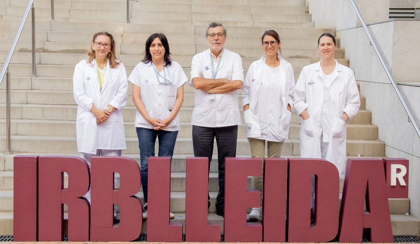
Dr. Xavier Matias-Guiu
Scientific manager
fjmatiasguiu.lleida.ics@gencat.cat
973 70 53 12
Dr. Maria Santacana
Technical manager
msantacana@irblleida.cat
973 70 53 12
Dr. Judit Pallarés
Collaborating facultative staff
jpallares.lleida.ics@gencat.cat
973 70 53 12
Dr. Sònia Gatius
Collaborating facultative staff
sgatius.lleida.ics@gencat.cat
973 70 53 12
Maria Carrelé
Technical staff collaborator
mcarrele@irblleida.cat
973 70 53 12